Peripheral Retinal Pathology Including Lattice Degeneration, Retinoschisis, and Masses
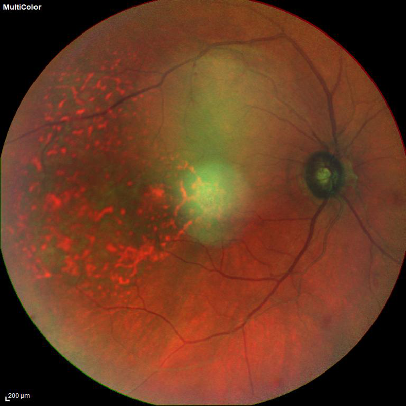 fundus photo of a choroidal tumor
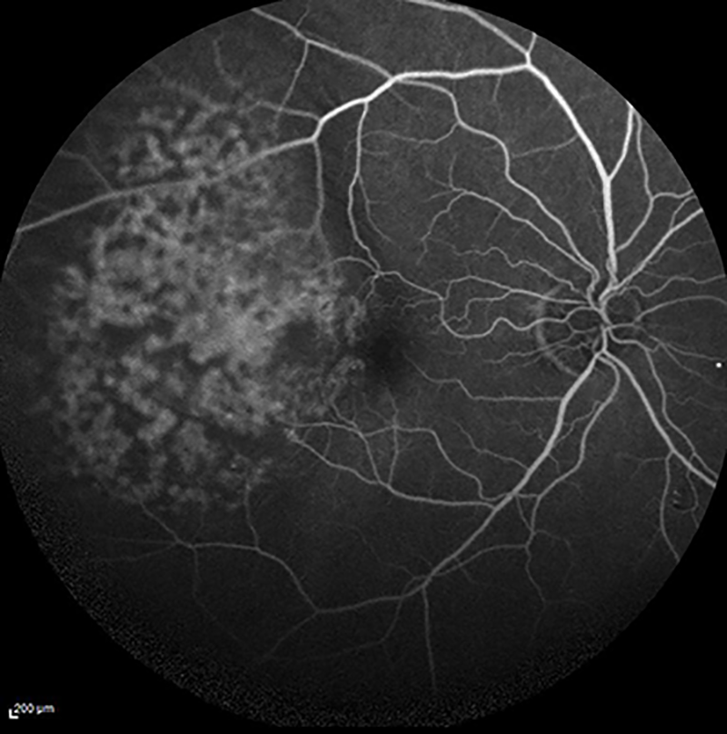 fluorescein angiogram of a choroidal tumor
The retina is divided into two main areas: the macula and the “peripheral” or surrounding retina. The macula is in the center of the retina and controls our detailed color vision. The peripheral retina makes up the rest of the retina and is responsible for our side and night vision. Lattice Degeneration, Retinoschisis, and Masses are just three of the different pathologies (or abnormal findings) that can be found in the peripheral retina.
What is Lattice Degeneration?
Lattice degeneration occurs when areas of the peripheral become thinner than normal. These areas of the retina are weaker and more prone to developing tears or holes, which can develop into retinal detachments. This condition is more common in patients who are nearsighted (cannot clearly see things that are far away). Often lattice degeneration can be present without any symptoms at all, but common symptoms are floaters and flashing lights. Lattice degeneration can be treated with a laser treatment (usually done in the office) which seals down the areas around the weakened retinal tissue. This prevents the development of retinal detachment in the future. Depending on your case, you may be recommended laser treatment or yearly observation for the treatment of lattice degeneration.
What is Retinoschisis?
Retinoschisis occurs when the retina separates into two layers. On examination, it can appear similar to a retinal detachment. It can technically occur in both the macula and the peripheral retina, however depending on where it occurs your treatment may differ. When the “schisis” or splitting occurs in the peripheral retina, it can usually be monitored to make sure it does not spread to include the macula, or central retina. In rare instances, retinoschisis can progress into the macula or develop into a combined retinal detachment with retinoschisis. At that time surgical intervention is recommended.
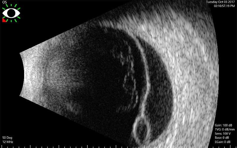
ultrasound of a retinal detachment with retinoschisis
Tumors of the eye:
A tumor can be described as an abnormal growth of tissue in the eye. This tissue may be benign (or non-cancerous and does not spread to other tissues) or malignant (cancerous, may spread to other structures). There are many different types of tumors that can form in the eye; a few examples are: choroidal nevus (see entry above for more information), choroidal (uveal) melanoma, hemangioma, retinoblastoma, and tumors that have spread from cancerous cells in other parts of the body, most commonly breast, lung, or prostate cancers. If your retinal specialist finds that you have a tumor in the back of your eye, you may be referred to another eye specialist, an ocular oncologist, who may take you through a series of specialized tests and procedures to diagnose your condition.
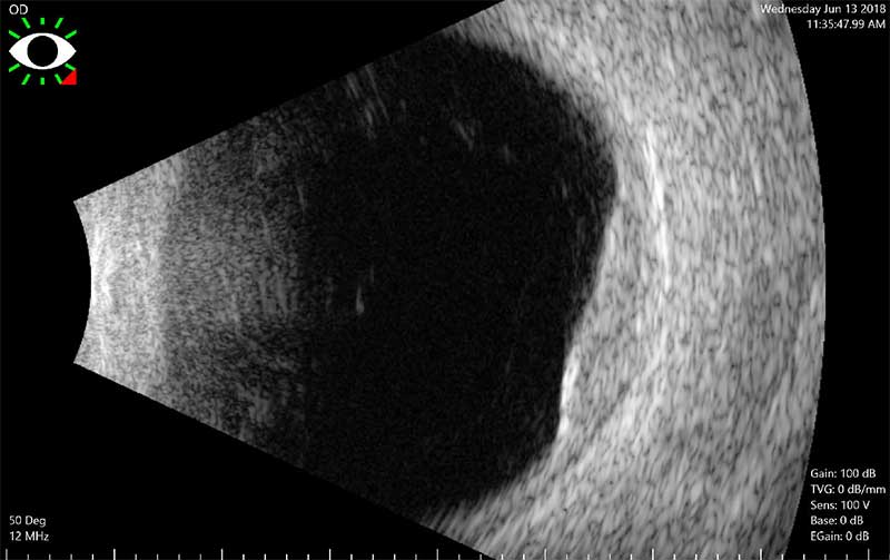
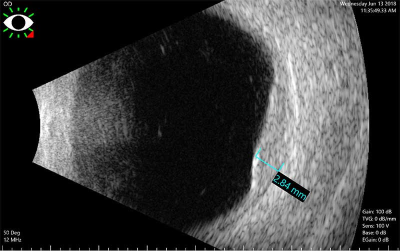
ultrasounds of a choroidal metastasis tumor