Macular Hole
What is a Macular Hole?
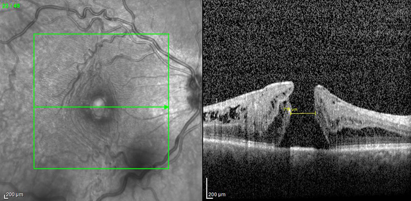
 OCT of a macular hole
A macular hole occurs when a tear or opening develops in the macula. The macula is a specific part of the retina that is responsible for your detailed vision. It is located at the center of the retina. This hole in the macula causes vision to be distorted or blurry.
What are the causes of a Macular Hole?
A macular hole can occur when portions of the vitreous (jelly that fills the inside of the eye) pull away from the macula. If enough traction is used, a hole or opening may form in the macula. Age most commonly contributes to the formation of a macular hole. Also, women around the age of 50 or 60 more commonly develop macular holes for unclear reasons. Additionally, macular holes can form due to eye injuries and swelling from other eye diseases.
How are Macular Holes diagnosed?
In order to diagnose this condition, you will undergo a detailed eye exam after your pupils have been dilated with eyedrops. Your retina specialist will then use a special lens to look at the inside of your eye. Special pictures of the eye may be taken, with a special technique called Optical Coherence Tomography, which captures detailed images of the back of your eye. Sometimes special dye test may be used as well, such as a fluorescein angiogram.
How is this condition treated?
Typically, the most effective treatment of a macular hole is a vitrectomy surgery. This involves removing vitreous from the eye and replacing it with a gas bubble. After having a gas bubble placed inside the eye, you will be required to maintain your face pointing down for several days. This position will cause the gas bubble to press against the hole, allowing it to close itself. Over the process of 2-8 weeks the bubble will slowly be absorbed by the body and be replaced with a natural fluid that the eye produces. Successful closure of the macular hole with vitrectomy is generally very high, approximately 95-99%, depending on the specific case. Once the hole closes, the vision typically slowly improves and distortion decreases. The visual outcome typically depends on the size of the hole and the length of time it was present before surgery. Typically, vision does not return all the way to normal, but patients often note that they see less distortion and are able to read a few more lines on the eye chart. Please remember that, as with any surgical procedure, there are risks involved with a vitrectomy, which you should discuss with your retinal surgeon.
Restrictions?
There are a few restrictions for patients after surgery to decrease chances of procedure failure and ensure eye health is maintained. While the gas bubble is in the eye, you cannot travel to higher elevations, fly in an airplane, or have specific types of anesthesia, such as nitrous oxide (“laughing gas;” commonly used by dentists). Any of these activities can lead to severe pain and possible blindness. Your retina specialist will tell you when it is safe to resume these activities.