Choroidal Melanoma


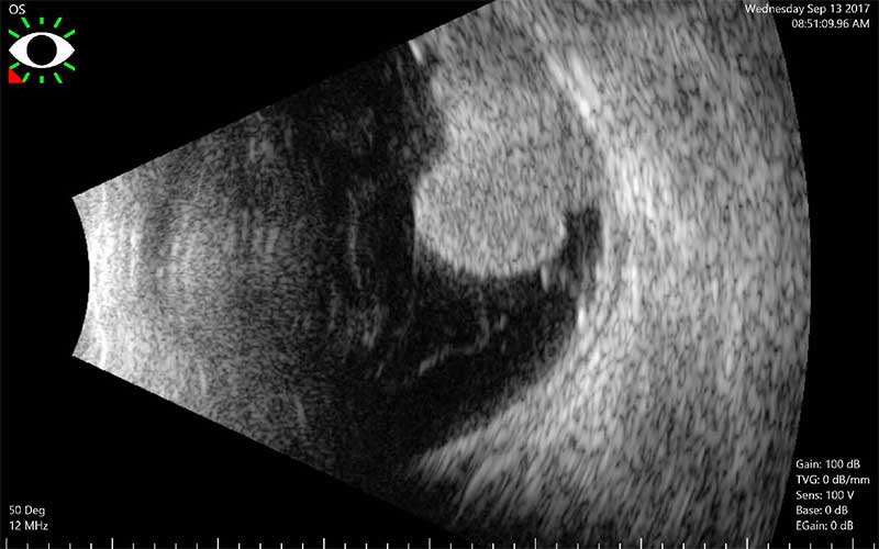
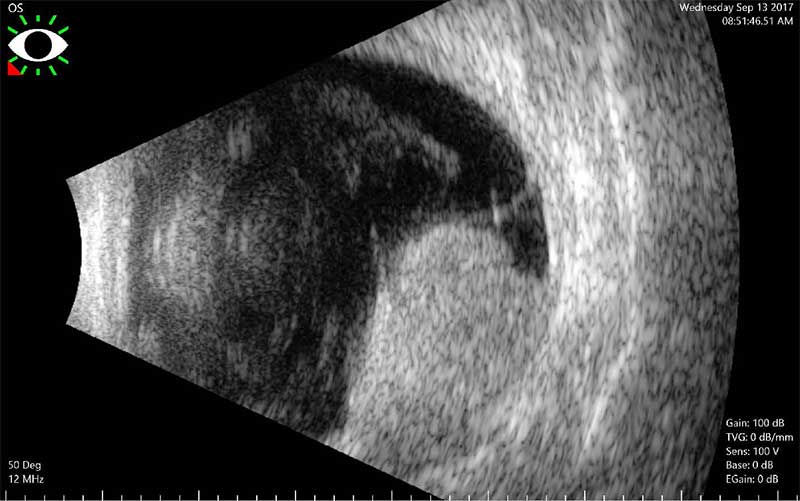
ultrasounds of a choroidal melanoma
What is a choroidal melanoma?
A melanoma is a type of cancer that comes from special pigment-producing cells, called melanocytes. When a melanoma occurs inside a part of the eye called the choroid, it is called a choroidal melanoma. (Note, melanoma can occur anywhere inside the body, as well as various parts of the eye).
Am I at risk for having a Choroidal Melanoma?
While choroidal melanomas are generally rare, they are the most common type of cancer that effects the eyes of adults. We do not know what causes choroidal melanomas, but studies have shown that middle-age Caucasians, who are exposed to increased amounts of UV light, seem to be at higher risk. Sometimes, occupational exposure can increase the risk for developing this condition. For instance, arc welders have a slightly increased risk.
What are the symptoms of a Choroidal Melanoma?
Depending on where in the eye the melanoma grows, patients can range from having no symptoms at all to having blurred vision, floaters or flashes of light. In rare occasions, some patients with this condition may also present with pain.

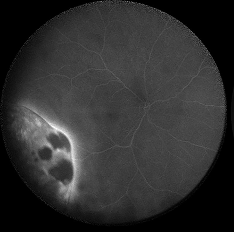
fluorescein angiogram of choroidal melanoma
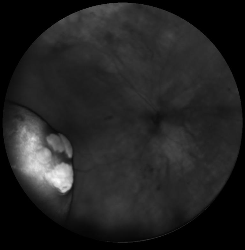
 ultra-wide field fundus photo of choroidal melanoma
How do you diagnose and treat a Choroidal Melanoma?
In order to diagnose a Choroidal Melanoma, you will undergo a dilated eye exam. Sometimes, your retina specialist will use specialized testing in order to confirm your diagnosis and fully evaluate how the melanoma affects the retina. For instance, a fluorescein angiogram (photographic dye test) can be used to look at blood vessels around the melanoma. OCT testing (optical coherence tomography) can also be used, which allows your doctor to evaluate a high-resolution picture of the retina so that the extent of damage can be assessed. Optical Ultrasound is used to measure the size of the tumor and look at its internal characteristics. Treatment depends on the size and location of the melanoma, and sometimes radiation or laser therapy may be used. Unfortunately, if the tumor is very large, the only treatment may be to remove the eye. Goals of treatment are to maintain as much vision as possible while controlling the size of the melanoma. Early treatment helps to reduce the risk of metastasis (spread to other areas of the body), which will help avoid any future serious health problems or death due to the melanoma.